Saturday, 12 July 2014
.gif)
Friday, 4 July 2014

Sunday, 29 June 2014
How to Pick a Good Watermelon AND 2 easy ways to cut one!
AFTAAAR WITHOUT WATERMELON .... No No No No
So how do I pick a good watermelon for my family to eat?
Taking direction from The Food Channel I look for these five things:
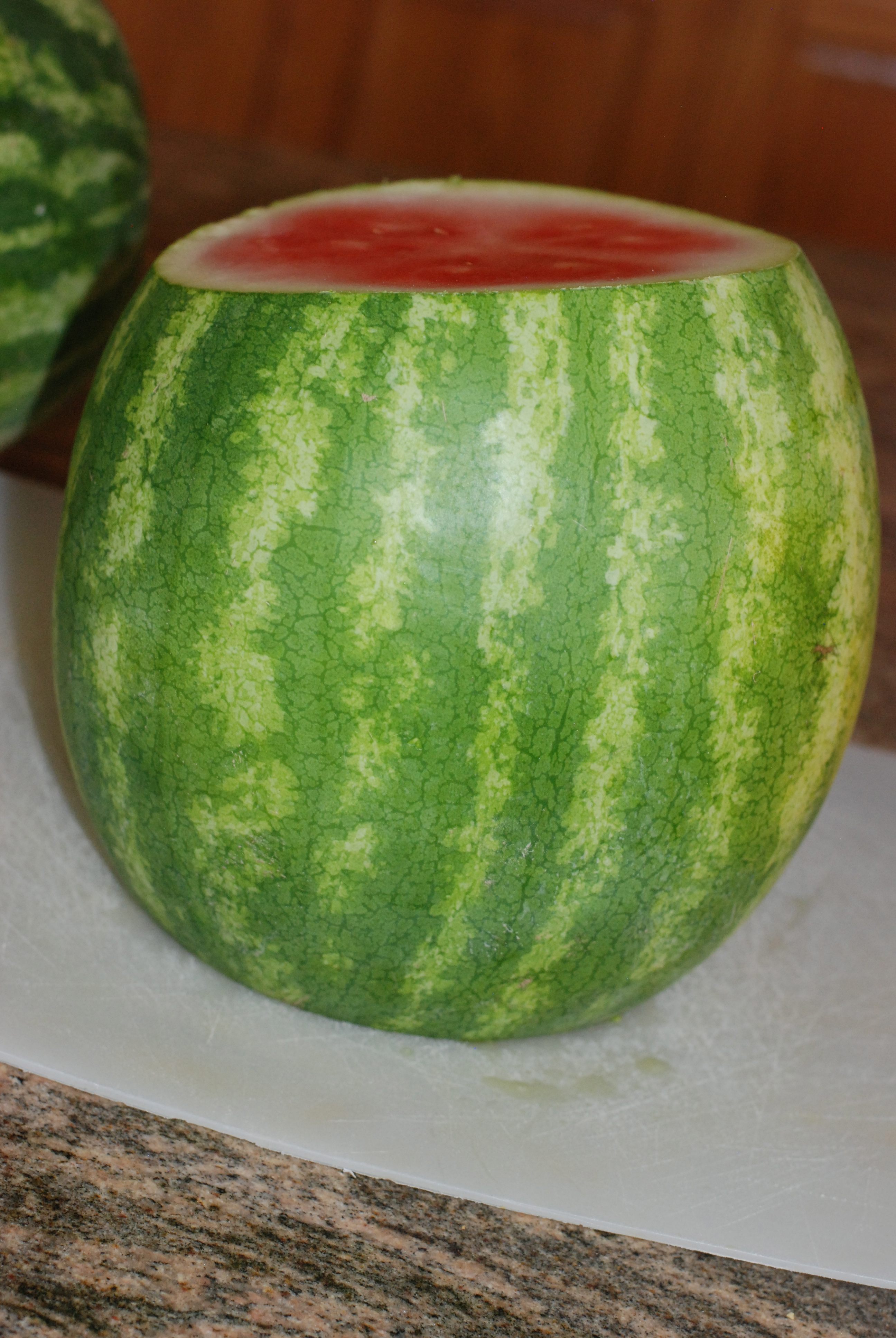
1. I look for watermelons that are nicely shaped and ones that feel firm. Make sure that they don’t have any cuts in them either.
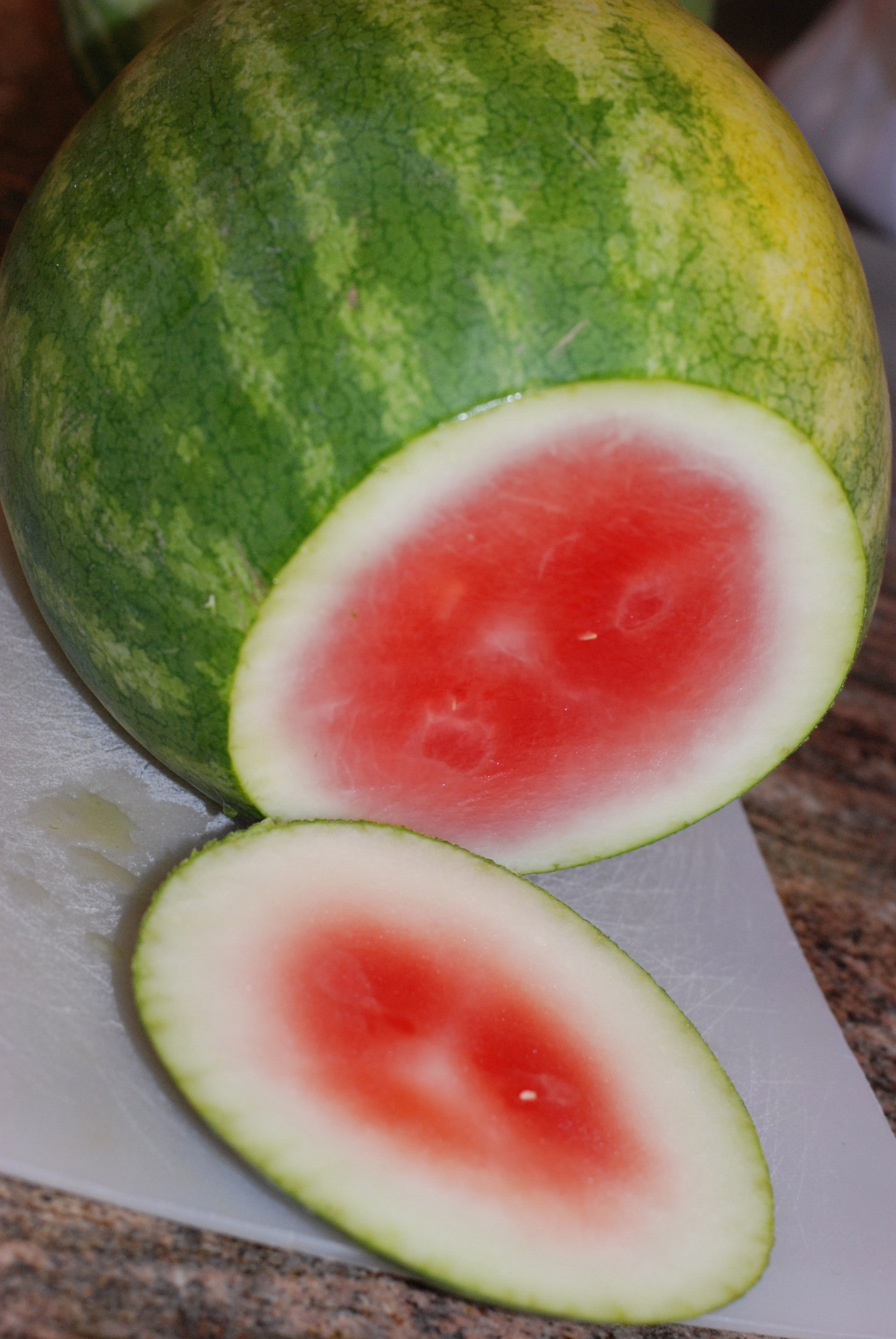
2. Pick your watermelon up to see if it feels “heavy”. Watermelons are 90% water, so the riper ones will have more water and weigh more
3. I look for watermelons that have a bright skin and vibrant green color
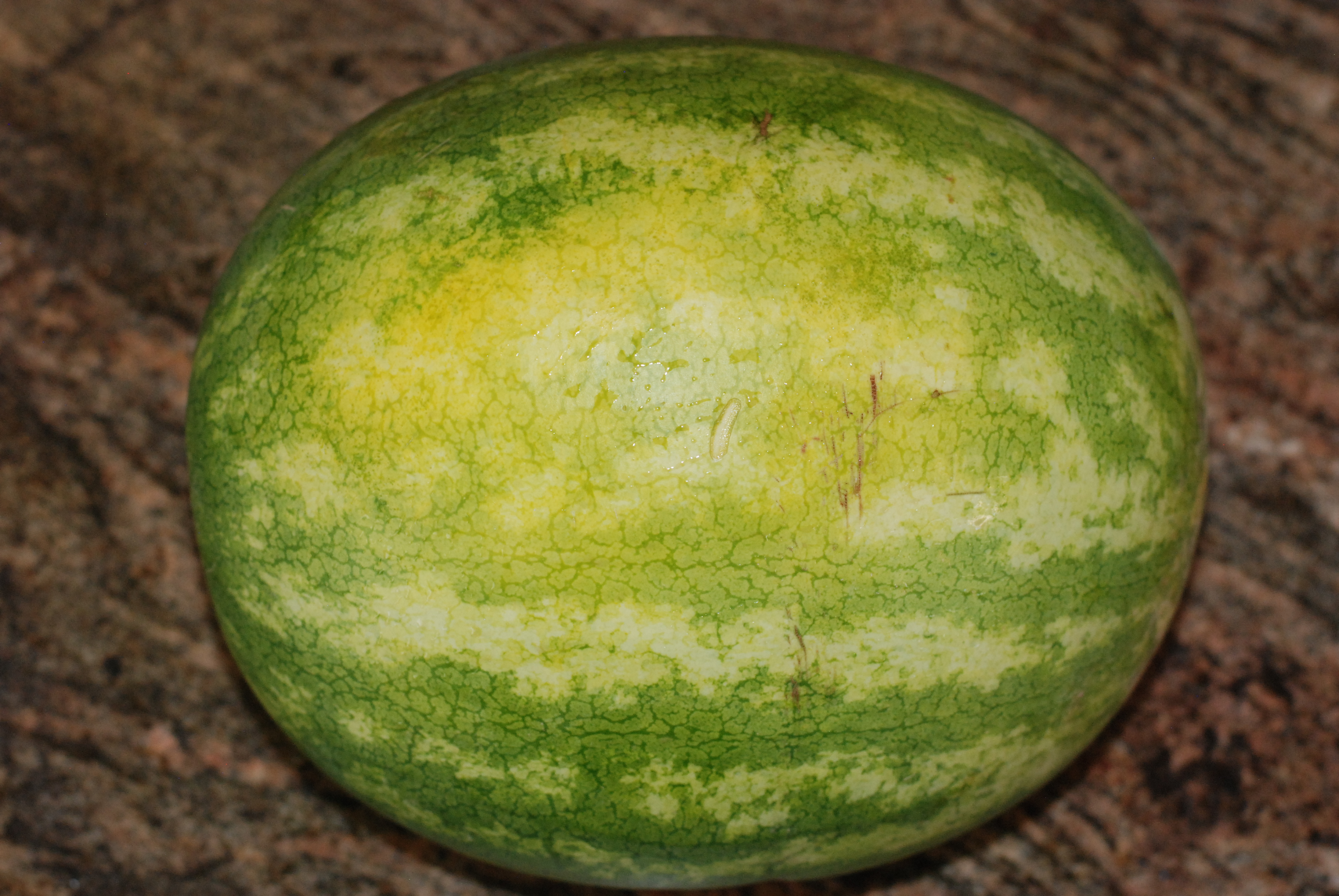
4. I used to not pick a watermelon that has a yellow side but have since found out that this yellow spot is a good thing and should be creamy looking. This is where it laid on the ground to ripen instead of being picked too early.
5. When “thumping” your watermelon, a solid sound means it’s not ripe enough, if it’s too ripe, it will sound thick but if it sounds hollow, that’s the one you want!
Below is a picture of the coveted “yellow spot”!
Now that you’ve found your perfect watermelon, how do you cut it?
Here are two EASY ways to get a beautifully cut watermelon!
The first method comes from a pin that I pinned from Fifteen Spatulas. First cut the bottoms off both ends of the watermelon:
Then stand it on one end:
Taking a knife, slice down and cut off the rind:

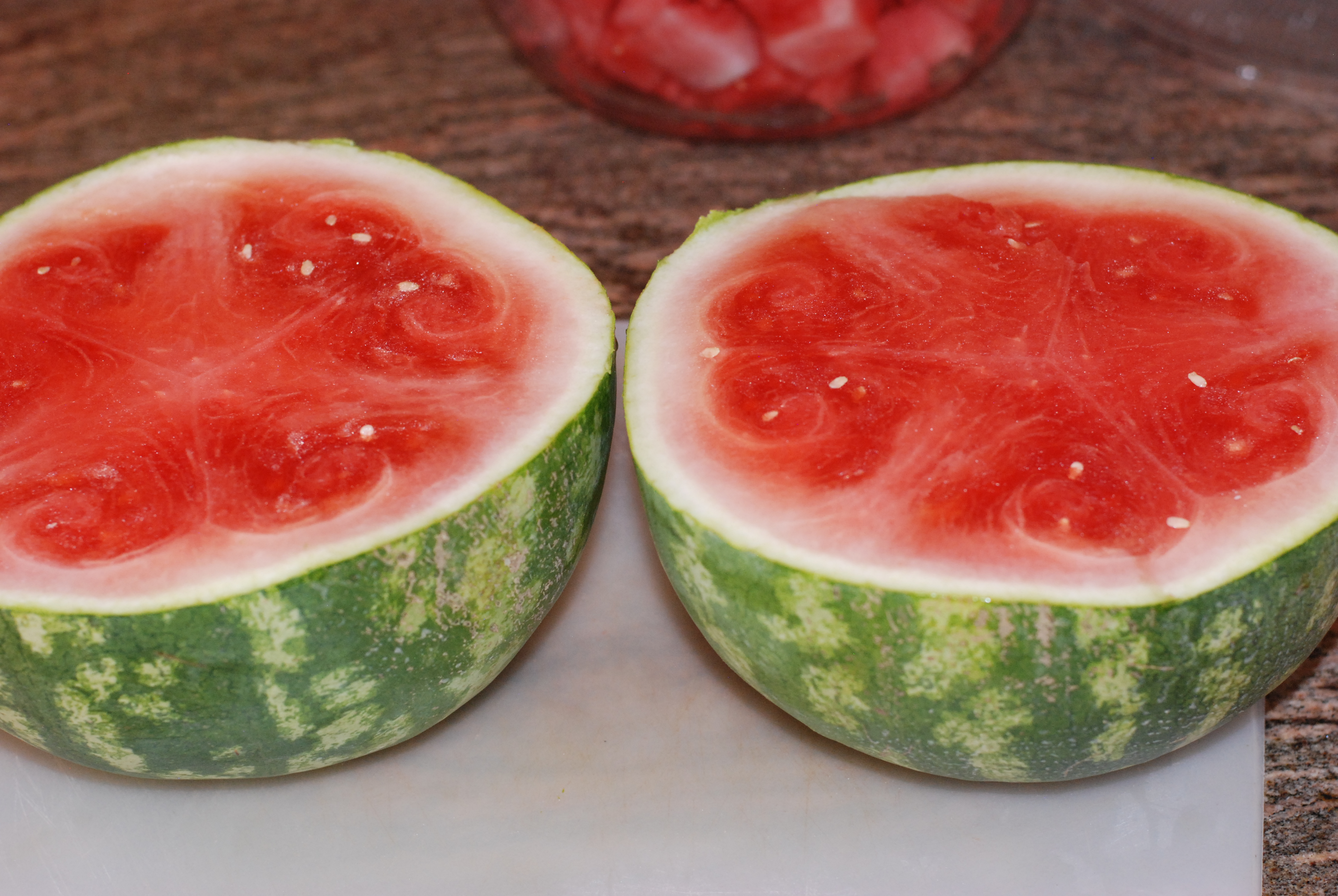
After removing all of the rind, cut into disks and then into strips and then into cubes:


Look how beautifully cubed the watermelon turned out!
Next up is a great way to cut watermelon if you have little bodies with little fingers as this takes out all the mess of eating sliced watermelon! This great idea comes from Mama Say What. The above method of cutting does a better job with larger melons. This next method works best for small to medium sized watermelons.
Cut your melon in half:
Lay the watermelon flat side down and cut into strips about 1 to 1/2 inches wide:

Then turn your watermelon and cut in the same size strips in the opposite direction:

What you will end up with are watermelon “strips” perfect for just picking up and eating!

Well, I’d love to sit around and eat watermelon with you (NOT) but my favorite son is calling.

Saturday, 28 June 2014
Tuesday, 24 June 2014

Wednesday, 18 June 2014

Saturday, 14 June 2014

Tuesday, 10 June 2014

Sunday, 8 June 2014
Friday, 6 June 2014
Sunday, 1 June 2014
Thursday, 29 May 2014

Monday, 26 May 2014
Friday, 23 May 2014

Sunday, 6 April 2014
Gram positive staining vs Gram negative staining
|
Gram positive staining
|
Gram negative staining
|
|
This is in contrast to Gram-negative, which cannot retain the crystal
violet stain, instead taking up the counterstain (safranin or fuchsine) and appearing red or pink
|
|
|
Gram-positive organisms
are able to retain the crystal violet stain because of the high amount of peptidoglycan in
the cell
wall.
|
|
|
Positive stain stick with
specimen and gives it's color
|
Negative dye doesn't stick
with the specimen but settle around it's outer boundary and forming a
silhouette. that negative stain produce a dark back ground around the cell
|
Importance of microbiology in nursing?
Q. Importance of
microbiology in nursing?
Microbiology has always been an essential and important
component of nursing and health science
curriculum. This is because of the relevance of microbiology in the hospitals
and our daily life, particularly in the areas of sterilization, cleaning,
aseptic processing, identification of infectious diseases, selection of drug
therapy, development of new drugs, preparation of vaccines, storage and
preservation of drugs. The importance of microbiology for nurses and health
professionals in the control and prevention of infection in hospital is more
and more recognized in recent years.
Basic knowledge of
microbiology is required in the field of nursing due to the following reasons:
1.
One must have an idea of how infections spread.
2.
Which surfaces are most susceptible to infectious
agents.
3.
How do you keep instruments aseptic and
contaminant-free.
4.
Recognize the symptoms of an infection.
5.
Recognize the type of infection at its early stage.
6.
How to carefully take care of an open wound without
infecting it.
7.
Recognize the type of infection as soon as it occurs.
8.
The nature of the organism and the factors affecting
its growth.
9.
The most susceptible means of disease transmission.
10. The composition
of chemicals, drugs, aseptic solution etc
11. The art of
working in a laboratory.
VIRUS
VIRUS
DEFINITION:
A virus (meaning a toxin or poision) is a small
infectious agent that replicates in the cell of
an organism.
The
causative agent of an infectious disease any of a large group of submicroscopic
infective agents that are regarded either as extremely simple microorganisms or
as extremely complex molecules, that are capable of growth and multiplication
only in living cells, and that cause various important diseases in humans,
animals, or plants.
VIRAL SIZES:
·
Smallest 0.02µm,
20 nanometers (polio virus)
·
Largest 0.3µm,
300 nanometers (smallpox virus)
HOST RANGE:
- Bunyaviruses: animals and plants
- Partitiviruses: plants and fungi
- Reoviruses: animals and plants
- Rhabdoviruses: animals and plants
- Phycodnaviruses: protozoa and plants
- Picornavirus-like viruses: plants and animals
- Totiviruses: protozoa / fungi and insects – tentative
CHARACTERISTICS OF VIRUS:
Living characteristics of viruses
a. They
reproduce at a fantastic rate, but only in living host cells.
Nonliving characteristics of viruses
a. They
are acellular, that is, they contain no cytoplasm or cellular organelles.
b. They
carry out no metabolism on their own and must replicate using the host cell's
metabolic machinery. In other words, viruses don't grow and divide. Instead,
new viral components are synthesized and assembled within the infected host
cell.
c. The
vast majority of viruses possess either DNA or RNA but not both.
GENERAL CHARACTERISTICS:
a. Viruses
are a cellular, non-cytoplasmic infectious agents.
b. They
are smaller than bacteria, and this can pass through bacteriological filter.
c. Viruses
are transmissible from disease to healthy organisms.
d. All
viruses are obligate parasites and can multiply only within the living host
cells.
e. Viruses
contain only a single type of nucleic acid either DNA or RNA.
f. Viruses
are host specific that they infect only a single species and definite cells of
the host organisms.
g. Viruses
are effective in very small doses. They are highly resistant to germicides and
extremes of physical conditions.
|
HELICAL VIRUS
|
Virus can be classified on following chracteristics:
Ø NUCLEIC
ACID:
·
ss DNA (single
stranded Deoxyribonucleic acid)
·
|
ENVELOPED
|
·
ss
RNA (single stranded ribonucleic acid)
·
ds RNA( double
stranded ribonucleic acid)
Ø MORPHOLOGY:
·
Helical
·
Polyhyderal
·
Enveloped
·
Complex
Ø REPLICATION:
·
Lytic cycle
·
Lysogenic cycle
STRUCTURE AND MORPHOLOGY:
HEAD: is protein membrane stuffed with molecule of
either DNA or RNA, consists of two parts CAPSID and ENVELOP.
COLLAR: base of the head, conecting head and tail.
HELICAL SHEATH: protein covering surrounding the
hollow core.
|
COMPLEX VIRUS
|
|
In the lysogenic cycle, the virus reproduces
by first injecting its genetic material, indicated by the red line, into
the host cell's genetic instructions
|
REPRODUCTION IN VIRUS:
Viruses uses their host cell to produce their
copies.
Virus replicates by two different methods:
Ø LYSOGENIC
CYCLE
Ø LYTIC
CYCLE
LYSOGENIC CYCLE
The lysogenic cycle is complementary to the
lytic cycle for viral entry and reproduction within cells. While the lytic
cycle is common to both animal viruses and bacterial phages, the lysogenic
cycle is more commonly found in animal viruses.
The following are the steps of the lysogenic
cycle:
1) Viral genome enters cell
2) Viral genome integrates into host cell
genome
3) Host cell DNA polymerase copies viral
chromosomes
4) Cell divides, and virus chromosomes are
transmitted to cell's daughter cells
5) At any moment when the virus is
"triggered", the viral genome detaches from the host cell's DNA and enters
stage 2 of the lytic cycle. While it is unclear as of yet what exactly
constitutes a "trigger" that activates the viral DNA from the latent
stage entered in Step 4, common symptoms that appear to "trigger" the
viral DNA are hormones, high stress levels (adrenaline), and free energy within
the infected cell.
An example of a virus that enter the lysogenic
cycle is herpes, which first enters the lytic cycle after infecting a human,
then the lysogenic cycle before travelling to the nervous system where it
resides in the nerve fibers as an episomal element. After a long period of time
(months to years) in a latent stage, the herpes virus is often reactivated to
the lytic stage during which it causes severe nervous system damage.
LYTIC CYCLE
lytic cycle is a viral replication cycle in which a virus
takes over a host cell's genetic material and uses the host cell's structures
and energy to replicate until the host cell bursts, killing it.
-A phage reproductive cycle that results in death of host cell.
-A phage reproductive cycle that results in death of host cell.
-A virulent phage: a
phage that reproduces only by a lytic cycle.
Step 1: A phage binds
its tail receptors to receptor cells on the outside of a cell.
Step 2: Part of its
tail contracts and allows the phage to enter the cell. The cell's DNA becomes
hydrolyzed.
Step 3: By using the
cell's resources, the phage produces proteins and copies of itself.
Step 4: Three separate
sets of proteins become individual phages.
Step 5: The phage
produces an enzyme that destroys the cell's bacterial wall and allows fluid to
enter. This causes the
cell to burst and release 100+ phage particles.
|
Figure
|
A generalized representation of
the replication of two viruses. Replication of a DNA virus is shown in (1);
replication of an RNA virus is displayed in (2).
|
Subscribe to:
Posts (Atom)